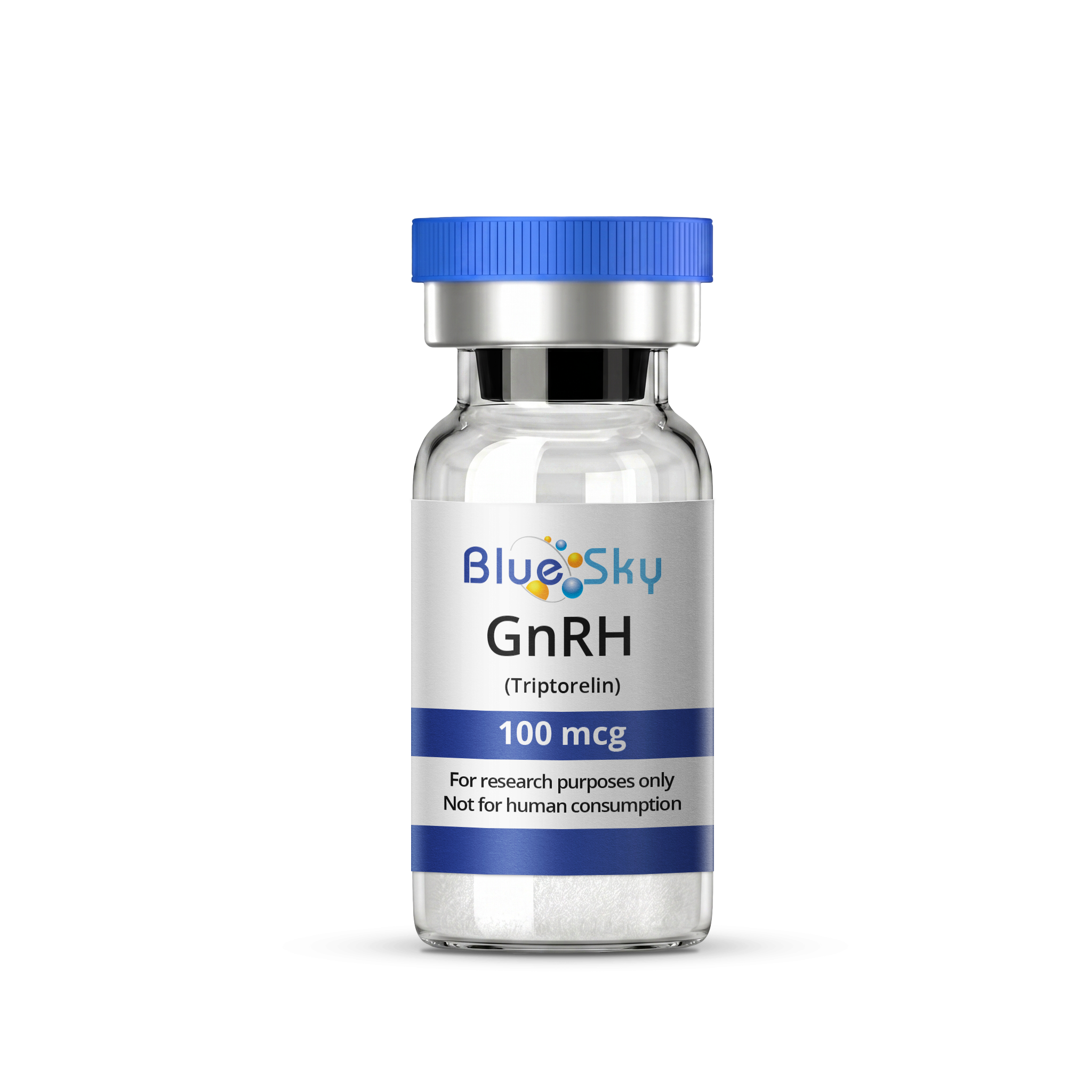
GnRH (Triptorelin) 100mcg

- Free Priority Shipping
- Orders over $75



Buy 1 Get 1 Free GnRH (Triptorelin) 100mcg :
| Unit Size | 100mcg/vial |
| Unit Quantity | 1 vial |
| Purity (Mass Spectrometry and UV) | 99.26% |
| Sequence | pGlu-His-Trp-Ser-Tyr-D-Trp-Leu-Arg-Pro-Gly-NH2 |
| Molecular Formula | C64H82N18O13 |
| Appearance | Lyophilized White Powder |
| Source | Chemical Synthesis |
| Storage | Lyophilized GnRH (Triptorelin) is stable at room temperature for 90 days. However, it is best stored in a freezer at -20°C for extended periods. After reconstitution, GnRH (Triptorelin) should be refrigerated at temperatures not exceeding 35°F. |
| Terms | The products we offer are intended for laboratory research use only. Please familiarize yourself with our terms of service prior to ordering. |
| COA Verification | Verify this COA on MZ Biolabs → |
GnRH (Triptorelin) 100mcg
View Research Overview
GnRH (Triptorelin) is a synthetic decapeptide analog of gonadotropin-releasing hormone (GnRH-I), a native ten-residue signaling peptide.
It differs from the native sequence by a single substitution: D-tryptophan replaces glycine at position 6. In laboratory research, it is studied in vitro as a reference agonist for the type I GnRH receptor, a class A G protein-coupled receptor (GPCR).
Peptide Structure and Chemistry
Triptorelin is ten residues long. It keeps two terminal features of the native peptide: a pyroglutamic acid (pGlu) N-terminus and an amidated glycine (Gly-NH2) C-terminus.
These blocked termini remove the free amine and free carboxyl groups that exopeptidases typically recognize, a common design feature in research peptides intended for stability in solution-based assays.
The defining modification is at position 6. Native GnRH carries glycine there, the only amino acid without a chiral center. Replacing it with the bulkier, D-configured tryptophan changes the peptide's preferred backbone geometry.
In GnRH peptide chemistry, a D-amino acid at position 6 is generally understood to stabilize a folded β-turn conformation that brings the two ends of the peptide closer together. This pre-organized shape is considered close to the receptor-bound form.
Primary Mechanism of Action
In vitro, triptorelin acts as an agonist at the type I GnRH receptor. Binding occurs in a pocket formed by the receptor's transmembrane helices and extracellular loops, with both termini of the folded peptide contributing to receptor contact.
Agonist binding is understood to shift the arrangement of the transmembrane helices, opening the intracellular surface of the receptor for G protein coupling.
The receptor couples primarily to Gαq/11. Downstream signaling studied in cell-based assays includes:
- Phospholipase C-β activation
- Inositol phosphate generation
- Intracellular calcium mobilization
- Diacylglycerol-dependent protein kinase C activation
- MAPK/ERK pathway signaling
A distinctive feature of the mammalian type I GnRH receptor is that it lacks a cytoplasmic C-terminal tail, including the helix 8 segment found in most class A GPCRs [1].
In many GPCRs, this tail region is associated with rapid desensitization and internalization. Its absence makes the GnRH receptor a useful model for studying sustained receptor signaling in cell-based systems.
In Vitro Research Findings
In cell-based receptor pharmacology, triptorelin is widely used as a reference agonist for the type I GnRH receptor. Its high receptor affinity and well-characterized structure make it a common benchmark in structure–activity studies of GnRH analogs.
Typical in vitro assay formats in which it serves as a reference compound include:
- Competitive radioligand binding in receptor-expressing cell lines
- Inositol phosphate accumulation assays
- Intracellular calcium flux measurements
- Site-directed mutagenesis studies mapping receptor binding residues
Comparisons between native GnRH and position-6-substituted analogs have been central to understanding how ligand conformation influences receptor binding. The bulky aromatic side chain of D-tryptophan is considered to add hydrophobic contact within the binding pocket, beyond its role in stabilizing the folded backbone.
Structural biology has added a further layer. A crystal structure of the GnRH receptor, solved with a small-molecule ligand, showed the receptor's N-terminus contributing to an enlarged binding pocket [1]. It serves as a structural reference for peptide-docking and receptor-modeling studies involving analogs such as triptorelin.
Chemical Stability in Aqueous Solution
Aqueous degradation of triptorelin has been characterized alongside gonadorelin and related analogs [2, 3]. These studies are directly relevant to laboratories preparing peptide solutions.
The L-serine at position 4 was identified as a key point of instability. It participates in solvent-catalyzed backbone hydrolysis and in hydroxide-catalyzed epimerization, the conversion of L-serine to its D-form [3].
Because epimerization is hydroxide-catalyzed, alkaline conditions are a known risk for solution stability. Comparable reactions proceeded with broadly similar kinetics across the analogs studied [3].
Epimerization also matters analytically. The resulting diastereomer has the same mass as the intact peptide, so mass spectrometry alone may not distinguish it. Chromatographic separation is needed to detect it.
Analytical Purity Verification
Triptorelin batches are typically verified by reversed-phase HPLC with UV detection for purity, combined with mass spectrometry for identity. For this peptide, analytical checks commonly look for:
- Truncated or deletion sequences from synthesis
- Incompletely deprotected byproducts
- The serine-4 epimer, which requires HPLC separation
- Hydrolysis fragments from cleavage near serine-4
The average molecular weight of the free base is approximately 1311.45 g/mol. The monoisotopic mass, approximately 1310.63 Da, is the reference value used when interpreting high-resolution mass spectrometry data. Material in acetate salt form carries a higher formula weight.
Storage and Handling in the Laboratory Setting
Lyophilized triptorelin is far more stable than material in solution. Laboratories typically keep vials frozen and sealed, and allow them to reach room temperature before opening to limit moisture uptake.
Once reconstituted, the serine-4 hydrolysis and epimerization pathways become active [3]. Common laboratory practice is to prepare solutions close to use, keep them refrigerated, avoid alkaline buffers, and aliquot to limit freeze–thaw cycles.
The tryptophan residues at positions 3 and 6 are susceptible to oxidation, so protection from light and prolonged air exposure is also standard practice.
References
- Yan W, Cheng L, Wang W, et al. Structure of the human gonadotropin-releasing hormone receptor GnRH1R reveals an unusual ligand binding mode. Nat Commun. 2020;11. doi:10.1038/s41467-020-19109-w
- Hoitink MA, Beijnen JH, Boschma MUS, et al. Identification of the degradation products of gonadorelin and three analogues in aqueous solution. Anal Chem. 1997;69:4972–4978.
- Hoitink MA, Beijnen JH, Boschma MUS, et al. Degradation kinetics of three gonadorelin analogues: developing a method for calculating epimerization parameters. Pharm Res. 1998;15:1449–1455. doi:10.1023/A:1011966024778
Important Notice
GnRH (Triptorelin) is provided exclusively for laboratory research purposes (in vitro). This peptide is not intended for human consumption or use, and any application outside of controlled laboratory research is prohibited. These findings do not establish safety, efficacy, or suitability for any human application, and have not been evaluated by the FDA or any regulatory agency.
Product Information
GnRH (Triptorelin) is supplied as a lyophilized powder for laboratory research use only.
 Oxytocin 5mg$31.17
 MELANOTAN II 10mg$32.47
 IGF-1 LR3 1mg$85.77
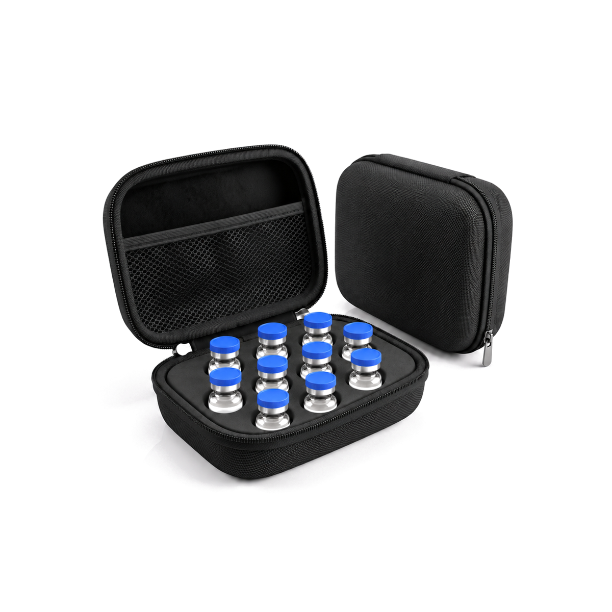 Storage Case (10)$9.99


 Login