January 2016

  1. CJC-1295: Super-GRF?

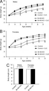
    Graphs demonstrating the ability of CJC-1295 to reverse M3-receptor knockout- (Br-M3-KO) induced effects on pituitary function in mice. From: Gautam D, Jeon J, Starost MF, et al. Neuronal M(3) muscarinic acetylcholine receptors are essential for somatotroph proliferation and normal somatic growth. Proceedings of the National Academy of Sciences of the United States of America. 2009;106(15):6398-6403, available through PNAS Open Access.

    Graphs demonstrating the ability of CJC-1295 to reverse M3-receptor knockout- (Br-M3-KO) induced effects on pituitary function in mice. From: Gautam D, Jeon J, Starost MF, et al. Neuronal M(3) muscarinic acetylcholine receptors are essential for somatotroph proliferation and normal somatic growth. Proceedings of the National Academy of Sciences of the United States of America. 2009;106(15):6398-6403, available through PNAS Open Access.

    Introduction  CJC-1295 is an analog of the human growth-hormone releasing factor (also known as GHRH)1,2. This synthetic peptide has selectivity for albumin (through a covalent bond with its 34th amino acid residue), thus increasing its half-life once administered2. Such a property confers advantages for CJC-1295 as a research compound and possible future therapeutic agent over human GHRH. CJC-1295 has demonstrated the ability to increase the AUC for human growth hormone (hGH) that was four-fold compared to that elicited by hGHRH. Its activity can be detected in laboratory animals for a number of days after administration2. The administration of CJC-1295 may also result in the increased release of insulin-like growth factor 1 (IGF-1)1. It may also have a positive effect on the proliferation of somatotroph cells1. CJC-1295 possesses the distinguishing feature of a modified lysine residue at the C-terminus2.  CJC-1295 and Growth Promotion  Treatment with CJC-1295 may restore growth rates to nearly normal in animal models of growth hormone deficiency. A study compared the effects of 2μg subcutaneous CJC1295 given once every 24, 48 or 72 hours for five weeks in week-old GH-knockout mice1. Similar groups of knockout and normal mice injected once daily with saline were used as controls. Knockout mice who received 2μg CJC-1295 every 24 hours exhibited significant increases in bodyweight and length compared to those injected every 48 or 72 hours, or to knockout controls1. The weights and lengths of the '24 hour' group were not significantly different from those of the normal control mice1. These growth-related values observed in the '48-' and '72-' hour groups were also significantly different to those in the control knockout group1. The '24 hour' group exhibited a 13-fold increase in pituitary development (as measured by total RNA detection) compared to knockout control mice1. This group also had an eleven-fold increase in pituitary GH expression (as indicated by mRNA detection) compared to knockout controls1. In addition, the '24-hour' group had significantly greater concentrations of IGF-1 compared to normal controls1. This indicates the potential of CJC-1295 to affect growth and somatotroph proliferation, provided the dose used is administered in line with the half-life of albumin (approximately 24 hours in mice)2.  CJC-1295 and the Neuronal Regulation of the Pituitary  This compound may also have a role in the determination of the nerve-cell activity (e.g. which receptors are involved, etc.) underpinning the normal function of the pituitary gland, which is not as yet fully understood. For example, it is not clear how the cells of the anterior pituitary are regulated, although muscarinic acetylcholine receptors appear to be involved in this3. Animals that do not express the M3 subtype of this receptor in brain cells exhibit a dwarfed phenotype with significantly reduced GH release and thus impaired anterior pituitary development and function4. 'Knockout' mice were treated with 2μg subcutaneous CJC-1295 daily for eight weeks, with similarly-treated normal mice as controls4. Body length and anterior pituitary size were not significantly different between these two groups4. GH and IGF-1 concentrations in the CJC-1295-treated knockout mice were increased at least two-fold compared to identical untreated knockout controls4. This supports the theory that neurons associated with the release of GHRH in the brain express M3 muscarinic receptors. References: 1. Alba M, Fintini D, Sagazio A, et al. Once-daily administration of CJC-1295, a long-acting growth hormone-releasing hormone (GHRH) analog, normalizes growth in the GHRH knockout mouse. American Journal of Physiology - Endocrinology and Metabolism. 2006;291(6):E1290-E1294. 2. Jette L, Leger R, Thibaudeau K, et al. Human growth hormone-releasing factor (hGRF)1-29-albumin bioconjugates activate the GRF receptor on the anterior pituitary in rats: identification of CJC-1295 as a long-lasting GRF analog. Endocrinology. 2005;146(7):3052-3058. 3. Giustina A, Veldhuis JD. Pathophysiology of the neuroregulation of growth hormone secretion in experimental animals and the human. Endocrine reviews. 1998;19(6):717-797. 4. Gautam D, Jeon J, Starost MF, et al. Neuronal M(3) muscarinic acetylcholine receptors are essential for somatotroph proliferation and normal somatic growth. Proceedings of the National Academy of Sciences of the United States of America. 2009;106(15):6398-6403.
  2. Albuterol and the Study of Lung Function and Reactivity

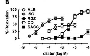
    Albuterol (ALB) in comparison with a novel bronchodilator (RGZ) in the relaxation of isolated mouse airway tissue. From: Donovan C, Simoons M, Esposito J, Ni Cheong J, Fitzpatrick M, Bourke JE. Rosiglitazone is a superior bronchodilator compared to chloroquine and beta-adrenoceptor agonists in mouse lung slices. Respiratory research. 2014;15:29, reproduced under the terms of a Creative Commons Attribution 4.0 International License

    Albuterol (ALB) in comparison with a novel bronchodilator (RGZ) in the relaxation of isolated mouse airway tissue. From: Donovan C, Simoons M, Esposito J, Ni Cheong J, Fitzpatrick M, Bourke JE. Rosiglitazone is a superior bronchodilator compared to chloroquine and beta-adrenoceptor agonists in mouse lung slices. Respiratory research. 2014;15:29, reproduced under the terms of a Creative Commons Attribution 4.0 International License

    Introduction Albuterol is historically linked to bronchodilation, as it activates the beta-2 subtype of the adrenergic receptor (β2-AR). It is available as two discrete isomers; S-albuterol and R-albuterol (also known as albuterol). A treatment consisting of a mixture of these isomers may have a complementary effect on therapeutic corticosteroids and a beneficial effect on inflammation, but purified S-albuterol lacks this property1. Albuterol elicits generally beneficial effects in lung tissue through the modulation of different ions, occurring as a result of β2-AR activation. However, these same effects may precipitate into adverse events when albuterol is introduced into different tissues that also possess adrenergic receptors.  Albuterol and Cardiovascular Effects  High-dose albuterol is associated with increased risks of tachycardia and tachypnea (accelerated heart rate and breathing) and with ventricular arrythmia. This may be associated with sharp decreases in cellular potassium as a result of β2-AR activation, which may lead to increased weakness in various muscle tissue types2. These effects may result in acute myocardial injury in severe cases2. On the other hand, albuterol may also increase intracellular calcium in these cells, which may be associated with increased contractility in cardiac muscle3. Albuterol and Respiratory Conditions Treatment with albuterol is a well-established method in the alleviation of conditions such as asthma. Albuterol at concentrations of 107M to 106M significantly reduced the contraction in isolated guinea pig trachea mediated by 107M to 103M insulin4. The administration of this compound may also address conditions such as acute respiratory distress syndrome (ARDS) and other acute lung conditions through the regulation of sodium/potassium-ATPase (Na/K-ATPase)5. This enzyme is involved in the reduction of Na+ concentrations in alveolar spaces, thus contributing to the control of fluid accumulation and swelling in these areas of lung tissue5. Recent research has improved the understanding of how albuterol regulates this ATPase. A study using rat alveolar cells demonstrated the ability of the molecule to induce the influx of intracellular calcium through calcium release-activated calcium (CRAC) channels. This in turn enhances the aggregation of Na/K-ATPase at the plasma membrane of alveolar cells, which is also mediated by β2-AR activation5. Albuterol may also be useful in trials that determine the genes that may be involved in respiratory function and health (e.g. airway responsiveness or the regulation of inflammation). The administration of albuterol failed to change lung resistance in mice with an extra copy of the Plp gene, although it had a negative effect on this measure in normal and carrier mice6. This indicates a role for the gene in responsiveness. Similarly, albuterol may also be used in the study of conditions that may be associated with increased risks of respiratory dysfunction. For example, congenital cryptorchidism (or retention of the testicles in the abdominal cavity) may be comorbid with asthma symptoms in some species7. Treatment with albuterol significantly reduced methacholine-resistance in both rats with this condition and corresponding control animals7. This treatment also resulted in the increased down-regulation of interleukin-4 and -6 in the lungs of rats affected by cryptorchidism7. This indicates a role for albuterol in the control of respiratory inflammation for animals with co-morbid conditions. Albuterol may also be used in trials assessing novel treatments for airway constriction and other respiratory symptoms8.  References: 1. Ameredes BT, Calhoun WJ. Levalbuterol versus albuterol. Current allergy and asthma reports. 2009;9(5):401-409. 2. Matos J, Jenni S, Fischer N, Bienz H, Glaus T. [Myocardial damage and paroxysmal ventricular tachycardia in a dog after Albuterol intoxication]. Schweizer Archiv fur Tierheilkunde. 2012;154(7):302-305. 3. Ogrodnik J, Niggli E. Increased Ca(2+) leak and spatiotemporal coherence of Ca(2+) release in cardiomyocytes during beta-adrenergic stimulation. The Journal of physiology. 2010;588(Pt 1):225-242. 4. Sharif M, Khan BT, Ajmal K, Anwar MA. Acute effect of insulin on guinea pig airways and its amelioration by pre-treatment with salbutamol. JPMA. The Journal of the Pakistan Medical Association. 2014;64(8):932-935. 5. Keller MJ, Lecuona E, Prakriya M, et al. Calcium release-activated calcium (CRAC) channels mediate the beta(2)-adrenergic regulation of Na,K-ATPase. FEBS letters. 2014;588(24):4686-4693. 6. Rodriguez E, Sakowski L, Hobson GM, et al. Plp1 gene duplication inhibits airway responsiveness and induces lung inflammation. Pulmonary pharmacology & therapeutics. 2015;30:22-31. 7. Rodriguez E, Barthold JS, Kreiger PA, et al. The orl rat is more responsive to methacholine challenge than wild type. Pulmonary pharmacology & therapeutics. 2014;29(2):199-208. 8. Donovan C, Simoons M, Esposito J, Ni Cheong J, Fitzpatrick M, Bourke JE. Rosiglitazone is a superior bronchodilator compared to chloroquine and beta-adrenoceptor agonists in mouse lung slices. Respiratory research. 2014;15:29.
  3. Epithalon: A short aside in the research of cancer and aging?

    The pineal gland in cross-section, from which epithalon was originally derived.

    The pineal gland in cross-section, from which epithalon was originally derived. "Pineal gland - high mag" by Nephron - Own work. Licensed under CC BY-SA 3.0 via Commons -

    Introduction Epithalon (also referred to as epitalon or AEDG) is a small peptide made of four amino acids (i.e. a sequence of Ala-Glu-Asp-Gly). Some researchers, particularly those involved in the isolation of this tetrapeptide, claim that epithalon confers beneficial effects in the areas of cancer prevention, the treatment of age-related physiological changes and nerve cell activity1. Epithalon may have some advantages as a research peptide, mostly due to its relatively small mass. However, its role in the management of states such as tumorigenesis and aging is uncertain at best.  Epithalon and its purported effects in Cancer Development Some scientists have claimed that epithalon may prevent tumor development in animal models of carcinogenesis. A study used an inbred mouse strain to assess this property2. 61 female mice were treated with 0.5μg/ml subcutaneous epithalon five days a week for 26 weeks. The number of spontaneously-forming tumors in these mice was compared to those in 56 identical mice receiving saline as a control. The mice receiving epithalon developed fewer tumors compared to controls, and exhibited no metastases compared to three in the control mice2. However, no significant differences were reported here2. Treatment with epithalon was shown to have no effect in the rate of malignant lymphoma in another study using a mouse line susceptible to this condition3. Other researchers have claimed that 0.1μg epithalon given five times a week prevented spontaneous tumor formation in rats living in ambient environmental light, but not those exposed to standard laboratory or constant illumination4. Epithalon and Aging Aging in animals is characterized by an increased susceptibility to DNA damage, (e.g. mutations) shorter telomeres (which essentially measure the ability of the cell to replicate its DNA in the course of proliferation) and other deficiencies in normal processes that protect cells from damage and death. There are some reports claiming evidence that epithalon may conserve cell survival through the restoration of telomere and telomerase structure and function, which degrade as a cell ages5. The authors of another study claimed a significant (20%) decrease in the chromosomal aberration (another marker of aging) of mice carrying mutations that promote aging (SAMP-1 mice) compared to identical untreated controls6. Another study compared female SAMR-1 and SAMP-1 mice treated with either melatonin (a neurotransmitter that is also linked to increased protection against aging and age-related physiological irregularities) or epithalon five times weekly for four weeks to untreated control mice (although the group size was unreported)3. These authors observed no difference between the treated or control animals of either strain in metabolic parameters. However, they claimed that both epithalon and melatonin alleviated irregularities in the estrous cycle of the treated animals compared to controls3. The last 10% of the SAMP-1 mice to die exhibited an increased rate of aging and a decreased mean life span compared to SAMR-1 mice, although treatment with epithalon or melatonin resulted in increased mean and maximum lifespan in these SAMP-1 mice compared to their untreated controls3. A study of aging under different lighting conditions claimed that, while treatment with 0.1μg epithalon five times a week did not affect the survival of rats living under standard laboratory illumination regimens, it did increase the maximum survival times of rats living under either constant or ambient (i.e. natural to the surrounding environment) illumination by 24 and 95 days respectively4. References: 1. Vanhee C, Moens G, Van Hoeck E, Deconinck E, De Beer JO. Identification of the small research tetra peptide Epitalon, assumed to be a potential treatment for cancer, old age and Retinitis Pigmentosa in two illegal pharmaceutical preparations. Drug testing and analysis. 2015;7(3):259-264. 2. Kossoy G, Anisimov VN, Ben-Hur H, Kossoy N, Zusman I. Effect of the synthetic pineal peptide epitalon on spontaneous carcinogenesis in female C3H/He mice. In vivo (Athens, Greece). 2006;20(2):253-257. 3. Anisimov VN, Popovich IG, Zabezhinskii MA, et al. [Effect of epitalon and melatonin on life span and spontaneous carcinogenesis in senescence accelerated mice (SAM)]. Voprosy onkologii. 2005;51(1):93-98. 4. Vinogradova IA, Bukalev AV, Zabezhinski MA, Semenchenko AV, Khavinson V, Anisimov VN. Effect of Ala-Glu-Asp-Gly peptide on life span and development of spontaneous tumors in female rats exposed to different illumination regimes. Bulletin of experimental biology and medicine. 2007;144(6):825-830. 5. Khavinson V, Bondarev IE, Butyugov AA. Epithalon peptide induces telomerase activity and telomere elongation in human somatic cells. Bulletin of experimental biology and medicine. 2003;135(6):590-592. 6. Rosenfeld SV, Togo EF, Mikheev VS, Popovich IG, Khavinson V, Anisimov VN. Effect of epithalon on the incidence of chromosome aberrations in senescence-accelerated mice. Bulletin of experimental biology and medicine. 2002;133(3):274-276.

Items 1 to 3 of 3 total