February 2015

  1. What is MGF Peptide?

    mgfpeptidegraph

    Growth rates of cultured rabbit mesenchymal stem cells in response to the administration of MGF peptide at the concentrations outlined in the legend. From Tong et al., Mechano-growth factor accelerates the proliferation and osteogenic differentiation of rabbit mesenchymal stem cells through the PI3K/AKT pathway. BMC Biochemistry. 2015;16(1):1.

      What is MGF Peptide?  Mechano-growth factor peptide (MGF peptide) is a 24-amino acid carboxy-terminal fragment of the insulin-like growth factor (IGF-1) protein1. It is a product of IGF-1 mRNA splicing2. The expression of MGF peptide is related to tissue damage and mechanical stimuli2. For example, the release of MGF peptide has been observed to increase in response to skeletal muscle injury3. This is thought to induce hypertrophy in damaged muscle tissue4. Some proteins found in muscle, such as titin and myomesin, are associated with the expression of this peptide in cultured murine myoblasts and myotubes2. The mechanical stimuli associated with the normal growth of tissues may also result in the expression of MGF peptide4. Other forms of stimulus that may be associated with the synthesis of MGF peptide are increased acidity and heat5. It has been linked to improvements in facets of muscular regeneration. These include delayed myoblast fusion and improved satellite cell proliferation1. MGF peptide has also been linked to protective roles against the death of nerve and heart muscle cells2. Therefore, this peptide may have a role in tissue growth and repair6. Studies involving MGF Peptide An in vitro trial using mouse myoblasts showed that MGF peptide at concentrations of up to 500ng/ml did not increase their proliferation or their differentiation into myotubes1. To confirm this, the researchers then incubated murine skeletal muscle stem cells with the peptide. No effects on the differentiation or numbers of these cells were observed1. On the other hand, MGF peptide has been associated with growth and differentiation in other cell and animal types. The exposure of rabbit mesenchymal stem cells to this peptide resulted in their growth and differentiation into osteoblasts7. This was mediated by the phosphorylation of mTOR and Akt7. (Conversely, the study as above found no increases in the phosphorylation of Akt in murine heart muscle cells, as claimed in previous reports1.) Another study using rat osteoblasts found that there was a significant increase in proliferation, and a slight decrease in differentiation, after three days of MGF peptide administration6. However, after chronic (three weeks) administration, improved differentiation of these cells was observed6. Mouse models of myocardial infarction may be characterized by a significant decline in diastolic and systolic hemodynamics as well as pathological hypertrophy within approximately ten weeks of an infarction8. The administration of MGF peptide within 12 hours of these events in mice had a positive effect on these hemodynamics, but not on hypertrophy8. MGF peptide administration for eight weeks following infarctions resulted in significant improvement in systolic function8. Another study assessed the expression of MGF peptide in the growth plates of piglets4. Porcine Mgf mRNA was found to be increased in the hypertrophic parts of growth plates4. However, the administration of exogenous MGF peptide to cultures of growth plate cells or chondrocytes did not result in proliferation4. A study of the effects of MGF peptide on rat mesenchymal stem cells found that this may enhance the stiffness and traction force associated with the migration of these cells9. The phosphorylation of ERK was associated with MGF peptide-induced migration9. Repetitive stretching in muscles of Wistar rats was associated with the significantly increased expression of Mgf mRNA, but only in the presence of the anabolic steroid metenolone10. References: 1.  Fornaro M, Hinken AC, Needle S, et al. Mechano-growth factor peptide, the COOH terminus of unprocessed insulin-like growth factor 1, has no apparent effect on myoblasts or primary muscle stem cells. American journal of physiology. Endocrinology and metabolism. 2014;306(2):E150-156. 2.  Kravchenko IV, Furalyov VA, Popov VO. Stimulation of mechano-growth factor expression by myofibrillar proteins in murine myoblasts and myotubes. Molecular and cellular biochemistry. 2012;363(1-2):347-355. 3.  Vassilakos G, Philippou A, Tsakiroglou P, Koutsilieris M. Biological activity of the e domain of the IGF-1Ec as addressed by synthetic peptides. Hormones (Athens, Greece). 2014;13(2):182-196. 4.  Schlegel W, Raimann A, Halbauer D, et al. Insulin-like growth factor I (IGF-1) Ec/Mechano Growth factor--a splice variant of IGF-1 within the growth plate. PloS one. 2013;8(10):e76133. 5.  Kravchenko IV, Furalyov VA, Popov VO. Hyperthermia and acidification stimulate mechano-growth factor synthesis in murine myoblasts and myotubes. Biochemical and biophysical research communications. 2008;375(2):271-274. 6.  Xin J, Wang Y, Wang Z, Lin F. Functional and transcriptomic analysis of the regulation of osteoblasts by mechano-growth factor E peptide. Biotechnology and applied biochemistry. 2014;61(2):193-201. 7.  Tong Y, Feng W, Wu Y, Lv H, Jia Y, Jiang D. Mechano-growth factor accelerates the proliferation and osteogenic differentiation of rabbit mesenchymal stem cells through the PI3K/AKT pathway. BMC biochemistry. 2015;16(1):1. 8.  Shioura K, Pena J, Goldspink P. Administration of a Synthetic Peptide Derived from the E-domain Region of Mechano-Growth Factor Delays Decompensation Following Myocardial Infarction. International journal of cardiovascular research. 2014;3(3):1000169. 9.  Wu J, Wu K, Lin F, et al. Mechano-growth factor induces migration of rat mesenchymal stem cells by altering its mechanical properties and activating ERK pathway. Biochemical and biophysical research communications. 2013;441(1):202-207. 10.  Ikeda S, Kamikawa Y, Ohwatashi A, Harada K, Yoshida A. The effect of anabolic steroid administration on passive stretching-induced expression of mechano-growth factor in skeletal muscle. TheScientificWorldJournal. 2013;2013:313605.
  2. Melanotan II?

    melanotanii

    - Chemical structure of Melanotan II by TheCook, made available under the terms of the Creative Commons Attribution-Share Alike 3.0 Unported license.

      What is Melanotan II? This is an analog of the alpha-melanocyte-stimulating hormone, and an agonist of the melanocortin receptor types 3 and 4 (MCR3 and MCR4)1,2. The melanocortin system in the central nervous system is associated with the regulation of social, emotional and food intake behaviors. It is also associated with a role in mediating the beneficial effects of leptin in rats with uncontrolled diabetes3. The MCR4 receptor is also associated with the regulation of bodyweight4. Defective MCR4 is associated with disorders of satiety and with early-onset obesity5. Studies of Melanotan II and its Functions Social Functions Melanotan II may be able to influence factors such as partner preference in monogamous animals. This molecule was found to enhance this behavior in prarie voles, but not in more polygamous meadow voles1. This was found to be associated with the activation of oxytocin-releasing neurons in the hypothalamus and the enhancement of oxytocin release by melanotan II1. Subcutaneous administration of 10 mg/kg melanotan II to males and female neonates of the same species for the first seven days of life resulted in reduced play-fighting in males compared to matched controls6. It also enhanced partner preference in the treated females in adulthood6. Metabolic and Cardiovascular Functions The administration of melanotan II in a rat model of diabetes did not appear to reduce hyperglycemia3. Intraperitoneal melanotan II was associated with rapid decreases in body temperature and energy expenditure4. This did not affect feeding, however4. The effect of melanotan II on body temperature was found to be independent of MCR4 receptor availability, and may be linked to activity at vasopressin V1a receptors instead4. The infusion of melanotan II into the medial preoptic nuclei of rat brains was associated with thermogenesis in brown adipose tissue2. The molecule was also found to be associated with the expression of proteins involved in lipolysis and lipogenesis2. These actions appear to be regulated by the dorsomedial hypothalamus2. Knockdown studies in rats indicates that AMP-activated protein kinase (AMPK) may be involved in the mediation of melanotan II activity7. The microinjection of melanotan II into the paraventricular nucleus of rat brains resulted in an increase in mean arterial pressure8. MCR3 and 4 antagonists reversed this effect8. Melanotan II also increased cAMP in the paraventricular nucleus8. Another study compared mice with MCR4 receptor deficiency to identical animals with selectively restored MCR4 in proopiomelanocortin neurons. (These are associated with the effects of leptin on energy homeostasis9. The knockout of PTP1B, the negative regulator of leptin in these cells, was associated with increased sensitivity to the effects of melanotan II on feeding, bodyweight and energy expenditure in KO mice compared to controls9.) This resulted in observations of increased bodyweight and decreased energy expenditure in the MCR4-deficient mice only, although feeding, blood pressure and heart rate were similar between the two groups10. This may further indicate the role of this receptor - and thus of melanotan II - in energy homeostasis and bodyweight control. References:  1. Modi ME, Inoue K, Barrett CE, et al. Melanocortin receptor agonists facilitate oxytocin-dependent partner preference formation in the prairie vole. Neuropsychopharmacology : official publication of the American College of Neuropsychopharmacology. 2015. 2. Monge-Roffarello B, Labbe SM, Lenglos C, et al. The medial preoptic nucleus as a site of the thermogenic and metabolic actions of melanotan II in male rats. American journal of physiology. Regulatory, integrative and comparative physiology. 2014;307(2):R158-166. 3. Meek TH, Matsen ME, Damian V, Cubelo A, Chua SC, Jr., Morton GJ. Role of melanocortin signaling in neuroendocrine and metabolic actions of leptin in male rats with uncontrolled diabetes. Endocrinology. 2014;155(11):4157-4167. 4. Xu Y, Kim ER, Fan S, et al. Profound and rapid reduction in body temperature induced by the melanocortin receptor agonists. Biochemical and biophysical research communications. 2014;451(2):184-189. 5. Fani L, Bak S, Delhanty P, van Rossum EF, van den Akker EL. The melanocortin-4 receptor as target for obesity treatment: a systematic review of emerging pharmacological therapeutic options. International journal of obesity (2005). 2014;38(2):163-169. 6. Barrett CE, Modi ME, Zhang BC, Walum H, Inoue K, Young LJ. Neonatal melanocortin receptor agonist treatment reduces play fighting and promotes adult attachment in prairie voles in a sex-dependent manner. Neuropharmacology. 2014;85:357-366. 7. Tanida M, Yamamoto N, Shibamoto T, Rahmouni K. Involvement of hypothalamic AMP-activated protein kinase in leptin-induced sympathetic nerve activation. PloS one. 2013;8(2):e56660. 8. Li P, Cui BP, Zhang LL, Sun HJ, Liu TY, Zhu GQ. Melanocortin 3/4 receptors in paraventricular nucleus modulate sympathetic outflow and blood pressure. Experimental physiology. 2013;98(2):435-443. 9. De Jonghe BC, Hayes MR, Zimmer DJ, Kanoski SE, Grill HJ, Bence KK. Food intake reductions and increases in energetic responses by hindbrain leptin and melanotan II are enhanced in mice with POMC-specific PTP1B deficiency. American journal of physiology. Endocrinology and metabolism. 2012;303(5):E644-651. 10. do Carmo JM, da Silva AA, Rushing JS, Pace B, Hall JE. Differential control of metabolic and cardiovascular functions by melanocortin-4 receptors in proopiomelanocortin neurons. American journal of physiology. Regulatory, integrative and comparative physiology. 2013;305(4):R359-368.
  3. What is Clenbuterol?

    clenbuterolgraph

    The change in muscle tension as a result of a 5-minute fatigue protocol, showing the differences in maximal force and 50% initial force (black dotted lines), as well as the resistance to fatigue (Tlim) between rat extensor digitorum longus (EDL) muscle treated with clenbuterol and that of controls. From Sirvent et al., Effects of chronic administration of clenbuterol on contractile properties and calcium homeostasis in rat extensor digitorum longus muscle. PloS one. 2014;9(6):e100281.

      What is Clenbuterol?  Clenbuterol is an adrenergic beta-2 receptor agonist associated with increases in skeletal muscle growth1. It is associated with the increase of fast-twitch glycolytic muscle fibers as opposed to slow-twitch oxidative fibers2. This results in rapid changes (or 'remodeling') in the structure of skeletal muscle. Recent animal studies have shown that chronic subcutaneous clenbuterol administration may not increase relevant characteristics of muscular activity such as specific maximal tetanic force and contractile efficiency3. Another study demonstrated that chronic administration did not change relative force in the muscles of male Wistar rats2. However, it was associated with an increase in absolute force2. Clenbuterol increases calpain (a serine protease) activity2. Chronic clenbuterol intake was also found to be associated with an imbalance in calcium (Ca2+) signaling and concentration in muscle fibers2. A study using rat soleus muscle fibers showed inhibitory effects on intracellular Ca2+ and action potential4. The latter was not reversed in the presence of a beta-2 antagonist, indicating clenbuterol may also have activity at alternative receptors4. Therefore, clenbuterol may change the kinetics of muscle contraction via indirect pathways based on the disruption of cellular Ca2+ homeostasis3. Clenbuterol may be associated with the increased risk of muscle fatigue2. The switch to fast-twitch fiber activity results in varying levels of hypertrophy (rapid growth) in different muscle types5. This may be due to the responses of different subtypes of calpain to this drug5. Some researchers argue that clenbuterol administration results in myotoxicity at high doses (approximately 1mg/kg per day)6. Clenbuterol is also associated with damage to cardiac muscle4. Other Functions and Applications of Clenbuterol  Adrenergic beta-receptors are also present in the central nervous system. Their specific roles in the brain may be elicited through the experimental application of clenbuterol. Intraperitoneal clenbuterol (at three different doses) reduced visuospatial learning discrepancies in a rat model of impaired cortical function (elicited by significantly reduced beta-receptor expression in the neocortex)7. The injection of clenbuterol into a brain region (the ventral bed nucleus of the stria terminalis) implicated in the stress-related use of cocaine reinstated this behavior in rats8. This indicates that beta-receptors may regulate the activation of the ventral tegmental area (another brain region) associated with cocaine-seeking8. The infusion of clenbuterol into the basolateral complex of the amygdala enhanced 'emotional' and 'aversive' memory in rats9. Some studies have indicated that beta-2 receptors are associated with the breakdown of glycogen in astrocytes, which supplies energy to neurons in the absence of dietary glucose10. A study using a mouse model of this found that clenbuterol administration did not influence this, but that beta-2 activation was associated with the protection of axons10. Clenbuterol may also have a therapeutic role in developmental disorders. The administration of clenbuterol resulted in the reduction of respiratory and motor co-ordination deficits in a mouse model of Rett syndrome (knockout of the Mecp2 gene)11. It was also associated with improvements in the survival and social recognition abilities of young male mice with this mutation11. Clenbuterol also improved cognition and anxiety in older female heterozygous mice11.  References: 1. Emery PW, Rothwell NJ, Stock MJ, Winter PD. Chronic effects of beta 2-adrenergic agonists on body composition and protein synthesis in the rat. Bioscience reports. 1984;4(1):83-91. 2. Sirvent P, Douillard A, Galbes O, et al. Effects of chronic administration of clenbuterol on contractile properties and calcium homeostasis in rat extensor digitorum longus muscle. PloS one. 2014;9(6):e100281. 3. Py G, Ramonatxo C, Sirvent P, et al. Chronic clenbuterol treatment compromises force production without directly altering skeletal muscle contractile machinery. The Journal of physiology. 2015. 4. Head SI, Ha TN. Acute inhibitory effects of clenbuterol on force, Ca(2)(+) transients and action potentials in rat soleus may not involve the beta(2)-adrenoceptor pathway. Clinical and experimental pharmacology & physiology. 2011;38(9):638-646. 5. Douillard A, Galbes O, Rossano B, et al. Time course in calpain activity and autolysis in slow and fast skeletal muscle during clenbuterol treatment. Canadian journal of physiology and pharmacology. 2011;89(2):117-125. 6. Burniston JG, McLean L, Beynon RJ, Goldspink DF. Anabolic effects of a non-myotoxic dose of the beta2-adrenergic receptor agonist clenbuterol on rat plantaris muscle. Muscle & nerve. 2007;35(2):217-223. 7. Saez-Briones P, Soto-Moyano R, Burgos H, et al. beta-Adrenoceptor stimulation restores frontal cortex plasticity and improves visuospatial performance in hidden-prenatally-malnourished young-adult rats. Neurobiology of learning and memory. 2014;119c:1-9. 8. Vranjkovic O, Gasser PJ, Gerndt CH, Baker DA, Mantsch JR. Stress-induced cocaine seeking requires a beta-2 adrenergic receptor-regulated pathway from the ventral bed nucleus of the stria terminalis that regulates CRF actions in the ventral tegmental area. The Journal of neuroscience : the official journal of the Society for Neuroscience. 2014;34(37):12504-12514. 9. McReynolds JR, Anderson KM, Donowho KM, McIntyre CK. Noradrenergic actions in the basolateral complex of the amygdala modulate Arc expression in hippocampal synapses and consolidation of aversive and non-aversive memory. Neurobiology of learning and memory. 2014;115:49-57. 10. Laureys G, Valentino M, Demol F, et al. beta(2)-adrenergic receptors protect axons during energetic stress but do not influence basal glio-axonal lactate shuttling in mouse white matter. Neuroscience. 2014;277:367-374. 11. Mellios N, Woodson J, Garcia RI, et al. beta2-Adrenergic receptor agonist ameliorates phenotypes and corrects microRNA-mediated IGF1 deficits in a mouse model of Rett syndrome. Proceedings of the National Academy of Sciences of the United States of America. 2014;111(27):9947-9952.
  4. GHRP-2: An overview of the current research

    GHRP-2

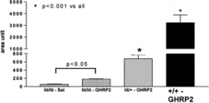
    Serum murine GH responses to 10 µg GHRP-2 (or saline (SAL)) in lit/lit (i.e. GHRH-deficient) mice in comparison to heterozygous or WT animals. From Peroni et al., Growth hormone response to growth hormone-releasing peptide-2 in growth hormone-deficient little mice. Clinics (Sao Paulo). 2012;67(3):265-272.


    What is GHRP-2?

    GHRP-2 is a small peptide (made of six amino acids) associated with the release of growth hormone (GH)1. This protein is associated with important roles in the regulation of sex hormones and metabolic control2. Decreases in this hormone are also associated with reductions in muscle mass, bone deterioration and possible cognitive deficits3. It is also linked to the increased risk of death from cardiovascular disease3. Growth hormone, as the name suggests, also plays a role in the growth and development of humans and other animals. GH deficiency has been shown to result in growth retardation.

     How does GHRP-2 act to increase GH?

    The release of GH is stimulated by the GH secretagogue (GHS) receptor, which is normally activated by ghrelin (also known as the GHS). GHRP-2 is a synthetic peptide that has also been shown to activate the GHS receptor (GHS-R1a)4. GHRP-2 (and ghrelin) also stimulates the release of prolactin, corticotropin and cortisol4. This has a positive effect on energy expenditure, appetite, heart muscle tone and sleep regulation. There is also a GH-releasing hormone (GHRH) present in humans and other animals, which stimulates the release of GH through its own receptor (GHRH-R). GHRP-2 complements this by potentiating the cAMP activation associated with GHRH-R binding, and may also promote the population of this receptor4. GHRP-2 has been found to significantly increase GH in genetically GHRH-deficient mice4. GHRP-2 is also associated with increases in IGF15.

    Other functions and uses of GHRP-2

    GHRP-2 may be administered orally. However, this is linked to reduced efficacy, as oral formulations have been found to result in poor absorption and thus reduced effects on (for example) GH secretion6. Intravenous solutions of GHRP-2 are regarded as the standard route, although subcutaneous and intraperitoneal administration is also used in some trials5-7. GHRP-2 may also have a potential role in analgesia (pain relief). This is due to recent observations that GHS-R1a activation by ghrelin in mice resulted in analgesia8. A recent study found that GHRP-2 also produced these effects. This was shown to be reversed by naloxone administration, indicating that GHS-R1a may be able to affect opioid receptors in the central nervous system8.

    GHRP-2 has also been found to bind to C36, which is a receptor for the pro-athergenic oxidized low-density lipoprotein5. It was found to reduce oxidative stress, but not plaque size, in a mouse model of atherosclerosis5. Subcutaneous GHRP-2 resulted in improvements of a rat model of acute lung injury9. This was attributed to reductions in TNFalpha, IL-6 and NFkappaB in the lung tissue of GHRP-2-treated rats9. This is further evidence of the anti-inflammatory properties of this peptide.

    Another study found that TNFalpha was also reduced by intraperitoneal GHRP-2 in a rat model of liver damage7. To confirm that this was as a result of exogenous GHRP-2, these researchers administered endotoxins and GHRP-2 to cultures of hepatocytes and nonparenchymal cells with hepatocytes. The peptide caused a reduction in TNFalpha mRNA in the latter, but not the former7. This indicates that GHRP-2 acts directly on the liver through nonparenchymal cells to reduce inflammation.

     References:

    1. Bowers CY. GH releasing peptides--structure and kinetics. J Pediatr Endocrinol. 1993;6(1):21-31.

    2. Veldhuis JD, Roemmich JN, Richmond EJ, Bowers CY. Somatotropic and gonadotropic axes linkages in infancy, childhood, and the puberty-adult transition. Endocr Rev. 2006;27(2):101-140.

    3. Norman C, Rollene NL, Erickson D, Miles JM, Bowers CY, Veldhuis JD. Estradiol regulates GH-releasing peptide's interactions with GH-releasing hormone and somatostatin in postmenopausal women. Eur J Endocrinol. 2014;170(1):121-129.

    4. Peroni CN, Hayashida CY, Nascimento N, et al. Growth hormone response to growth hormone-releasing peptide-2 in growth hormone-deficient little mice. Clinics (Sao Paulo). 2012;67(3):265-272.

    5. Titterington JS, Sukhanov S, Higashi Y, Vaughn C, Bowers C, Delafontaine P. Growth hormone-releasing peptide-2 suppresses vascular oxidative stress in ApoE-/- mice but does not reduce atherosclerosis. Endocrinology. 2009;150(12):5478-5487.

    6. Tanaka T, Hasegawa Y, Yokoya S, Nishi Y. Increased Secretion of Endogenous GH after Treatment with an Intranasal GH-releasing Peptide-2 Spray Does Not Promote Growth in Short Children with GH Deficiency. Clin Pediatr Endocrinol. 2014;23(4):107-114.

    7. Granado M, Martin AI, Lopez-Menduina M, Lopez-Calderon A, Villanua MA. GH-releasing peptide-2 administration prevents liver inflammatory response in endotoxemia. American journal of physiology. Endocrinology and metabolism. 2008;294(1):E131-141.

    8. Zeng P, Li S, Zheng Y-h, et al. Ghrelin receptor agonist, GHRP-2, produces antinociceptive effects at the supraspinal level via the opioid receptor in mice. Peptides. 2014;55:103-109.

    9. Li G, Li J, Zhou Q, Song X, Liang H, Huang L. Growth hormone releasing peptide-2, a ghrelin agonist, attenuates lipopolysaccharide-induced acute lung injury in rats. The Tohoku journal of experimental medicine. 2010;222(1):7-13. 

Items 1 to 4 of 4 total