1. The Importance of Bremelanotide Peptides for Laboratory Research Studies

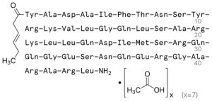
    Bremelanotide is a 7 amino acid (AA) analogue of α melanocyte-stimulating hormone (α-MSH) used in scientific research studies as a treatment for hyposexual sexual desire disorder (HSDD) in women1. As a peptide, bremelanotide is injected subcutaneously, although intranasal administration has shown efficacy in stimulating sexual arousal in female vitro test subjects. Male vitro test subjects with sexual dysfunction also demonstrate increased sexual arousal and erectile responses after subcutaneous bremelanotide injection, including subjects for whom Viagra was ineffective3. In addition to its beneficial role in sexual dysfunction studies, bremelanotide has excellent utility in many different fields of neuroscience research. DISCLAIMER: All products from Blue Sky Peptide are for laboratory research only and are not suitable for human consumption. Any other use violates the terms and conditions for purchase. Bremelanotide should only be used in carefully controlled laboratory conditions with appropriate personal protective equipment. If exposure occurs, immediately rinse the affected area and seek medical help if symptoms present. Bremelanotide is a melanocortin receptor (MCR) agonist that preferentially interacts with the melanocortin receptor subtype 4 (MC4R)4. The melanocortins and their receptors were first identified as pituitary factors involved in skin darkening in frogs. Eventually, however, the exploration of the melanocortin system extended to mammalian systems5. The melanocortin system contributes to skin pigmentation, inflammation, appetite, memory, and sexual behavior5-8. The different MCR subtypes play distinct roles and are differentially expressed across the body. Melanocortins, also known as melanocyte-stimulating hormones (MSH), are all small peptide hormones derived from proopiomelanocortin and serve as the primary agonists of MCRs along with adrenocorticotropic hormone (ACTH)9. MSH peptides come in 3 types: α-MSH, β-MSH, γ-MSH. 
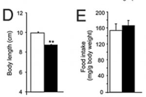
MC4R is primarily localized in the brain and is considered a neural MCR10. One of the primary roles of MC4R is regulating food intake by mediating the effects of leptin11. Administering α-MSH reduces feeding in obese rats, especially when applied directly to the paraventricular nucleus of the hypothalamus (PVN), a critical autonomic control center of the brain involved with stress responses, growth, and reproduction12-14. In addition to food intake, MC4R plays a role in other processes like glucose and lipid homeostasis, disease-associated lean body mass wasting, and cardiovascular function10. Reproductive behavior is also intrinsically tied to MC4R and its ligands. Mice with reduced or absent MC4R function exhibit several sexual dysfunctions15-17. Administration of MC4R agonist reversed these effects in some studies and caused erections and ejaculations in some animal models after central nervous system administration10,18. Considering the breadth of life-essential roles the melanocortin system has, an MC4R-specific agonist like bremelanocortin is valuable for many lines of neuroscience research. In addition, bremelanocortin is extensively characterized in the literature and is well-tolerated by many species, including rats and humans. Therefore, bremelanocortin is, and has been, a powerful research tool. References:
    1. Mayer D, Lynch SE. Bremelanotide: New Drug Approved for Treating Hypoactive Sexual Desire Disorder. Ann Pharmacother. 2020;54(7):684-690.
    2. Clayton AH, Althof SE, Kingsberg S, et al. Bremelanotide for female sexual dysfunctions in premenopausal women: a randomized, placebo-controlled dose-finding trial. Womens Health (Lond). 2016;12(3):325-337.
    3. Rosen RC, Diamond LE, Earle DC, Shadiack AM, Molinoff PB. Evaluation of the safety, pharmacokinetics and pharmacodynamic effects of subcutaneously administered PT-141, a melanocortin receptor agonist, in healthy male subjects and in patients with an inadequate response to Viagra. Int J Impot Res. 2004;16(2):135-142.
    4. Pfaus JG, Sadiq A, Spana C, Clayton AH. The neurobiology of bremelanotide for the treatment of hypoactive sexual desire disorder in premenopausal women. CNS Spectr. 2021:1-9.
    5. Wikberg JE, Muceniece R, Mandrika I, et al. New aspects on the melanocortins and their receptors. Pharmacol Res. 2000;42(5):393-420.
    6. Shen Y, Fu WY, Cheng EY, Fu AK, Ip NY. Melanocortin-4 receptor regulates hippocampal synaptic plasticity through a protein kinase A-dependent mechanism. J Neurosci. 2013;33(2):464-472.
    7. Catania A, Lonati C, Sordi A, Carlin A, Leonardi P, Gatti S. The melanocortin system in control of inflammation. ScientificWorldJournal. 2010;10:1840-1853.
    8. Ellacott KL, Cone RD. The role of the central melanocortin system in the regulation of food intake and energy homeostasis: lessons from mouse models. Philos Trans R Soc Lond B Biol Sci. 2006;361(1471):1265-1274.
    9. Gantz I, Fong TM. The melanocortin system. Am J Physiol Endocrinol Metab. 2003;284(3):E468-474.
    10. Tao YX. The melanocortin-4 receptor: physiology, pharmacology, and pathophysiology. Endocr Rev. 2010;31(4):506-543.
    11. Kask A, Rago L, Wikberg JE, Schioth HB. Evidence for involvement of the melanocortin MC4 receptor in the effects of leptin on food intake and body weight. Eur J Pharmacol. 1998;360(1):15-19.
    12. Ferguson AV, Latchford KJ, Samson WK. The paraventricular nucleus of the hypothalamus - a potential target for integrative treatment of autonomic dysfunction. Expert Opin Ther Targets. 2008;12(6):717-727.
    13. Hansen MJ, Ball MJ, Morris MJ. Enhanced inhibitory feeding response to alpha-melanocyte stimulating hormone in the diet-induced obese rat. Brain Res. 2001;892(1):130-137.
    14. Mountjoy KG, Mortrud MT, Low MJ, Simerly RB, Cone RD. Localization of the melanocortin-4 receptor (MC4-R) in neuroendocrine and autonomic control circuits in the brain. Mol Endocrinol. 1994;8(10):1298-1308.
    15. Sandrock M, Schulz A, Merkwitz C, Schoneberg T, Spanel-Borowski K, Ricken A. Reduction in corpora lutea number in obese melanocortin-4-receptor-deficient mice. Reprod Biol Endocrinol. 2009;7:24.
    16. Irani BG, Xiang Z, Moore MC, Mandel RJ, Haskell-Luevano C. Voluntary exercise delays monogenetic obesity and overcomes reproductive dysfunction of the melanocortin-4 receptor knockout mouse. Biochem Biophys Res Commun. 2005;326(3):638-644.
    17. Van der Ploeg LH, Martin WJ, Howard AD, et al. A role for the melanocortin 4 receptor in sexual function. Proc Natl Acad Sci U S A. 2002;99(17):11381-11386.
    18.       Bertolini A, Tacchi R, Vergoni AV. Brain effects of melanocortins. Pharmacol Res. 2009;59(1):13-47.
  2. How To Order Research Peptides Online

    Proteins and peptides are some of the most commonly used reagents in modern life sciences. From short-chain peptide receptor agonists to biotinylated-antibodies, peptide-based reagents play pivotal roles in every stage of research. The ubiquity of peptides has created a vibrant market for them and protein reagents with many suppliers at different levels. However, not all peptide suppliers are the same, and neither are their products. Therefore, the first step to ordering research peptides online is to identify whether a supplier’s products are of the exacting standards needed to conduct effective research. Bad reagents can ruin an experiment and potentially sink entire projects. More insidiously, research conducted with poorly tested components can get published and contribute to the growing reproducibility crisis in science1. In recent decades, irreproducibility has been brought to the forefront in science, with journals like Nature publishing extensively on the topic2,3. In 2015, one study estimated that up to 50% of preclinical research is irreplicable. This has cost approximately $28,000,000,000 in US dollars4. One key point made by some authors is that differences in reagents, especially protein products like antibodies, have made achieving reproducible results difficult. Therefore, identifying which protein and peptide products are validated is crucial. There are databases and websites in which antibodies from different manufacturers are listed and compared for efficacy, with links to studies that use them5. However, there is no such catalog or comparable resource for small peptide products6. This doesn’t mean that there is no way to be sure your peptide products are of the purity and quality you expect. Peptide products should undergo quality control analysis through multiple methods. These techniques include high-performance liquid chromatography (HPLC), mass spectrometry (MS), and nuclear magnetic resonance analysis (NMR)7-9. A reputable supplier will have quality control reports available on their product pages for some or all of these. Therefore, when shopping for research peptides online, it is pivotal to find quality control reports. Another factor to consider when shopping for research peptides online is customer service and accountability. Research is not a simple endeavor. Along the course of a project, it is a given that some experiments will not work out. In some cases, this is caused by the reagent. Even high-grade products will not work when used outside of their working conditions. By having direct contact with a peptide supplier, a researcher can troubleshoot issues that arise with their research peptide products. Additionally, a good return policy helps avoid losses if the product is not up to standards or does not match the application. Finally, and perhaps most obviously, pricing is critical. Research funds aren’t unlimited, and materials are only one of many costs that an investigator or lab manager must account for. Many suppliers offer scaling pricing options and periodic special offers. Additionally, some retailers have rewards programs that encourage repeat business. When searching for research peptides online, Blue Sky Peptides offers affordable peptides that come with quality control reports. We have in-depth customer support and a generous return policy. In addition, our site has regular sales, BOGO offers, discount codes, and our BSP Rewards Program. Visit our website or contact us today to see if our research peptides will meet your research needs. References:
    1. Williams M. Reagent Validation to Facilitate Experimental Reproducibility. Curr Protoc Pharmacol. 2018;81(1):e40.
    2. Baker M. 1,500 scientists lift the lid on reproducibility. Nature. 2016;533(7604):452-454.
    3. Begley CG, Ioannidis JP. Reproducibility in science: improving the standard for basic and preclinical research. Circ Res. 2015;116(1):116-126.
    4. Freedman LP, Cockburn IM, Simcoe TS. The Economics of Reproducibility in Preclinical Research. PLoS Biol. 2015;13(6):e1002165.
    5. Antibodypedia. https://www.antibodypedia.com/. Accessed August 28, 2021.
    6. Vergote V, Burvenich C, Van de Wiele C, De Spiegeleer B. Quality specifications for peptide drugs: a regulatory-pharmaceutical approach. J Pept Sci. 2009;15(11):697-710.
    7. Choules MP, Bisson J, Gao W, et al. Quality Control of Therapeutic Peptides by (1)H NMR HiFSA Sequencing. J Org Chem. 2019;84(6):3055-3073.
    8. Stalmans S, Gevaert B, Verbeke F, et al. Quality control of cationic cell-penetrating peptides. J Pharm Biomed Anal. 2016;117:289-297.
    9. Zeng K, Geerlof-Vidavisky I, Gucinski A, Jiang X, Boyne MT, 2nd. Liquid Chromatography-High Resolution Mass Spectrometry for Peptide Drug Quality Control. AAPS J. 2015;17(3):643-651.
  3. What Is Fragment 176-191 Peptide

    Fragment 176-191 is a small peptide that comprises the last 15 amino acids (AAs) from the c-terminal end of growth hormone (hGH)1. During childhood, hGH plays a critical role in growth regulation. However, in adulthood, hGH’s most well-known function is metabolic. Among other actions, hGH stimulates insulin-like growth factor-1 (IGF-1) production and release2-4. In addition, the release of hGH from the anterior pituitary gland is stimulated by hypothalamic growth hormone-releasing hormone (GHRH)5. Fragment 176-191 and similar hGH fragments have powerful lipolytic and anti-lipogenic properties that have drawn researchers’ interest for decades6,7. Early investigation showed that injecting rats with c-terminal fragments of hGH caused a temporary increase in blood glucose and a more persistent boost in plasma insulin. Interestingly, fragments that did not contain AAs 178-191 had no effect. This provided crucial early evidence suggesting a minimum required sequence for a peptide to be biologically active7. Later research found that both hGH and the slightly truncated fragment 177-191 could cause weight loss in mice by promoting lipolysis, although a previous report suggests that it inhibits lipogenesis instead8,9. Additionally, this study indicated that this is because the peptide sequence can boost the production of beta-3 adrenergic receptors (ADRB3)8. Interestingly, it does not appear that fragment 177-191 acts on hGH receptors and, unlike hGH, does not stimulate cell proliferation6. These results add to the body of evidence suggesting that hGH may be a prohormone with differentially truncated forms that have their own effects6,10. Finally, fragment 177-191 co-injected with hyaluronic acid (HA) reduced cartilage degeneration and the degree of motor disability in a rat osteoarthritis model11. These results highlight the utility of fragment 176-191 and related peptides as valuable research tools. Although a phase 2B clinical trial investigating fragment 177-191 as a treatment for obesity stalled, hGH c-terminal fragments provide means to manipulate lipid metabolism in the lab without the interfering factors of IGF-1 signaling alterations or GH receptor6,12. This specificity could one day help researchers uncover novel mechanisms and future therapies.  
    1. Wade JD, Ng FM, Bornstein J, Pullin CO, Pearce JS. Effect of C-terminal chain shortening on the insulin-antagonistic activity of human growth hormone 177--191. Acta Endocrinol (Copenh). 1982;101(1):10-14.
    2. Brinkman JE, Tariq MA, Leavitt L, Sharma S. Physiology, Growth Hormone. In: StatPearls. Treasure Island (FL)2021.
    3. Bidlingmaier M, Strasburger CJ. Growth hormone. Handb Exp Pharmacol. 2010(195):187-200.
    4. Ayuk J, Sheppard MC. Growth hormone and its disorders. Postgrad Med J. 2006;82(963):24-30.
    5. Vance ML. Growth-hormone-releasing hormone. Clin Chem. 1990;36(3):415-420.
    6. Heffernan MA, Thorburn AW, Fam B, et al. Increase of fat oxidation and weight loss in obese mice caused by chronic treatment with human growth hormone or a modified C-terminal fragment. Int J Obes Relat Metab Disord. 2001;25(10):1442-1449.
    7. Ng FM, Bornstein J. Hyperglycemic action of synthetic C-terminal fragments of human growth hormone. Am J Physiol. 1978;234(5):E521-526.
    8. Heffernan M, Summers RJ, Thorburn A, et al. The effects of human GH and its lipolytic fragment (AOD9604) on lipid metabolism following chronic treatment in obese mice and beta(3)-AR knock-out mice. Endocrinology. 2001;142(12):5182-5189.
    9. Wu Z, Ng FM. Antilipogenic action of synthetic C-terminal sequence 177-191 of human growth hormone. Biochem Mol Biol Int. 1993;30(1):187-196.
    10. Devesa J, Almenglo C, Devesa P. Multiple Effects of Growth Hormone in the Body: Is it Really the Hormone for Growth? Clin Med Insights Endocrinol Diabetes. 2016;9:47-71.
    11. Kwon DR, Park GY. Effect of Intra-articular Injection of AOD9604 with or without Hyaluronic Acid in Rabbit Osteoarthritis Model. Ann Clin Lab Sci. 2015;45(4):426-432.
    12. Heike Stier EV, David Kenley. Safety and Tolerability of the Hexadecapeptide AOD9604 in Humans. Journal of Endocrinology & Metabolism. 2013;3:7-15.
  4. What are Research Peptides

    Research peptides are small chains of amino acids used in pure and applied biological research but not in the clinic. Although there is overlap in the definitions of both peptides and proteins, peptides are generally defined as a small chain of between 2 and 50 amino acids (AAs)1. Peptides are further differentiated based on size. Single unbranched strands between 10-20 AAs are called polypeptides, and oligopeptides are greater than 20 AAs long1,2. Proteins are larger macromolecules consisting of hundreds to thousands of AAs that display secondary, tertiary, and quarternary structures1. Clinical and academic researchers have used research peptides to probe mechanisms underlying disease and as novel therapeutics. For example, one such study by Ahmed et al. investigated the effects of tatCN21 treatment in an animal model of global cerebral ischemia, the cessation or reduction of blood flow to the brain3. TatCN21 is a 21 AA peptide inhibitor of the protein calcium-calmodulin (CaM) dependent protein kinase IIα (CaMKIIα) that is attached to a “tat sequence,” which allows the peptide to be brought into the cell. Ahmed et al. found that treating rats with tatCN21 3 hours after GCI protected neurons against programmed cell death and reduced spatial memory deficits3. Another group investigated using a tat-conjugated peptide to block the interaction of Nuclear factor (erythroid-derived 2)-like 2 (Nrf2) and Kelch-like ECH-associated protein 1 (Keap1)4. Nrf2 is an important transcription factor that activates the cellular antioxidation response and regulates signaling pathways in cancer and inflammation5,6. Keap1 binds to Nrf2, preventing its activation and nuclear translocation 7. Steel et al. found that administering tat-conjugated peptide sequences that target this interaction could activate Nrf2 signaling and its downstream target genes in human THP-1 monocytes4. Many peptide hormones have been investigated for their potential beneficial properties in various diseases. Some of these, such as vasopressin, oxytocin, and insulin, have made it to the clinic, while others are in clinical trials8. Currently, 80 peptide medications are approved for medical use. However, bringing new peptides to market requires careful consideration of renal clearance and delivery methods to make sure the drugs make it to their target tissue9,10. Research peptides have significantly advanced our understanding in health and disease. However, quality research requires peptides you can trust. Blue Sky Peptide sells the highest-grade research peptides on the market. In addition, we regularly offer BOGO deals, weekly promotions, and a rewards program that gives back when you shop with us. Check out our product catalog or contact us to see what Blue Sky Peptide can do for your research projects. All products from Blue Sky Peptide are for laboratory research only and are not suitable for human consumption. Any other use violates the terms and conditions for purchase. References:
    1. Forbes J, Krishnamurthy K. Biochemistry, Peptide. In: StatPearls. Treasure Island (FL)2021.
    2. Friedberg F, Winnick T, Greenberg DM. Peptide synthesis in vivo. J Biol Chem. 1947;169(3):763.
    3. Ahmed ME, Dong Y, Lu Y, Tucker D, Wang R, Zhang Q. Beneficial Effects of a CaMKIIalpha Inhibitor TatCN21 Peptide in Global Cerebral Ischemia. J Mol Neurosci. 2017;61(1):42-51.
    4. Steel R, Cowan J, Payerne E, O'Connell MA, Searcey M. Anti-inflammatory Effect of a Cell-Penetrating Peptide Targeting the Nrf2/Keap1 Interaction. ACS Med Chem Lett. 2012;3(5):407-410.
    5. Kobayashi M, Yamamoto M. Molecular mechanisms activating the Nrf2-Keap1 pathway of antioxidant gene regulation. Antioxid Redox Signal. 2005;7(3-4):385-394.
    6. Moi P, Chan K, Asunis I, Cao A, Kan YW. Isolation of NF-E2-related factor 2 (Nrf2), a NF-E2-like basic leucine zipper transcriptional activator that binds to the tandem NF-E2/AP1 repeat of the beta-globin locus control region. Proc Natl Acad Sci U S A. 1994;91(21):9926-9930.
    7. Itoh K, Wakabayashi N, Katoh Y, et al. Keap1 represses nuclear activation of antioxidant responsive elements by Nrf2 through binding to the amino-terminal Neh2 domain. Genes Dev. 1999;13(1):76-86.
    8. Muttenthaler M, King GF, Adams DJ, Alewood PF. Trends in peptide drug discovery. Nat Rev Drug Discov. 2021;20(4):309-325.
    9. Mitragotri S, Burke PA, Langer R. Overcoming the challenges in administering biopharmaceuticals: formulation and delivery strategies. Nat Rev Drug Discov. 2014;13(9):655-672.
    10. Katz AI, Emmanouel DS. Metabolism of polypeptide hormones by the normal kidney and in uremia. Nephron. 1978;22(1-3):69-80.
  5. What is Fragment 176-191?

    The effects of impaired GRF release in mice (black bars) compared to healthy controls (white bars) on body length and feeding. Asterisks indicate significant difference compared to controls. From: Gautam D, Jeon J, Starost MF, et al. Neuronal M(3) muscarinic acetylcholine receptors are essential for somatotroph proliferation and normal somatic growth. Proceedings of the National Academy of Sciences of the United States of America. 2009;106(15):6398-6403, available through PNAS Open Access.

    The effects of impaired GRF release in mice (black bars) compared to healthy controls (white bars) on body length and feeding. Asterisks indicate significant difference compared to controls. From: Gautam D, Jeon J, Starost MF, et al. Neuronal M(3) muscarinic acetylcholine receptors are essential for somatotroph proliferation and normal somatic growth. Proceedings of the National Academy of Sciences of the United States of America. 2009;106(15):6398-6403, available through PNAS Open Access.

    Fragment 176-191 is a short peptide derived from human growth hormone (GH). It may also be regarded as an analog of the GH-releasing hormone, also known as GRF. This fragment is found at one end of the hormone’s peptide sequence, and is associated with some biochemical properties as a separate entity. At least twelve (178 to 190) of the amino acids in the sequence appear to be required for activity at the insulin receptor1. In this form, it may result in significant antagonism of this receptor, leading to insulin resistance in experimental animals1. Normally, growth hormone has a less detrimental effect on the effects of insulin, thus suggesting that glucose breakdown is promoted by the opposite end of the protein sequence2. Treatment with a peptide made of residues 177-191 of human GH may be associated with the inactivation of glycogen synthase phosphatase in rat muscle tissue, thus changing the concentration of active glycogen synthase in these cells3. Therefore, truncated forms of GH, including Fragment 177-191, may disrupt normal glucose or glycogen metabolism in vivo. Fragment 177-191 may also be responsible for reductions in the storage of fat molecules in rats, although it is not associated with the breakdown of existing fat stores5. The full, 16-amino-acid form of the fragment mimics growth-hormone releasing factor (GRF). GH- or GRF-analogs such as this may be associated with reduced adipose deposits in treated animals4. GRF and its analogs function to increase GH release from tissues such as the pituitary gland6. Therefore, they may also have knock-on effects on the concentrations of other hormones, such as progesterone6. GRF analogs may also be more stable than the full GH protein7. Fragment 176-191 may be used in studies investigating the putative roles of GH or GRF in processes such as aging and the response to changes in the feeding patterns of test animals8. It may also be used to further the understanding of the GH-GRF-IGF regulatory axis9. GRF is also thought to target cells in other parts of the body other than the pituitary in mammals9. In fact, it may have a role in perinatal and postnatal development9. It is a member of a superfamily of peptide hormones or hormone-like molecules that include PACAP, secretin, glucagon and glucagon-like proteins9. Therefore, a stable, bioactive GRF analog is a viable research reagent. GRF-releasing neurons are located in the hypothalamus, which implies another target for research incorporating Fragment 176-19110. These cells are still in need of study to elicit a full understanding of their function and their role in hormonal regulation. Fragment 176-191, as the name suggests, is a peptide fragment made of the 16 residues nearest the amino-terminus of human growth hormone. Therefore, it may elicit partial GH activity, while sacrificing other aspects of this. The fragment is also a GRF analog. Therefore, it may activate cells in the anterior pituitary, affect the levels of other hormones such as IGF-1 and potentially alter glucose metabolism in some animals. GRF analogs may also be useful in models of fat metabolism and storage.    References:
    1. Wade JD, Ng FM, Bornstein J, Pullin CO, Pearce JS. Effect of C-terminal chain shortening on the insulin-antagonistic activity of human growth hormone 177--191. Acta endocrinologica. 1982;101(1):10-14.
    2. Mondon CE, Reaven GM, Ling N, Lewis UJ, Frigeri LG. Amino-terminal peptide of growth hormone enhances insulin action in normal rats. Endocrinology. 1988;123(2):827-833.
    3. Macaulay SL, Armstrong JM, Bornstein J. Regulation of glycogen synthase activity in muscle by a C-terminal part sequence of human growth hormone. Archives of biochemistry and biophysics. 1983;224(1):365-371.
    4. Mangili A, Falutz J, Mamputu J-C, Stepanians M, Hayward B. Predictors of Treatment Response to Tesamorelin, a Growth Hormone-Releasing Factor Analog, in HIV-Infected Patients with Excess Abdominal Fat. PLoS ONE. 2015;10(10):e0140358.
    5. Wu Z, Ng FM. Antilipogenic action of synthetic C-terminal sequence 177-191 of human growth hormone. Biochemistry and molecular biology international. 1993;30(1):187-196.
    6. Haldar A, Prakash BS. Effects of growth hormone-releasing factor on growth hormone response, growth and feed conversion efficiency in buffalo heifers (Bubalus bubalis). Veterinary journal (London, England : 1997). 2007;174(2):384-389.
    7. Hu M, Tomlinson B. Growth hormone-releasing factor agonists for the treatment of HIV-associated lipodystrophy. Current opinion in investigational drugs (London, England : 2000). 2010;11(10):1143-1150.
    8. Sun LY, Spong A, Swindell WR, et al. Growth hormone-releasing hormone disruption extends lifespan and regulates response to caloric restriction in mice. eLife. 2013;2:e01098.
    9. Campbell RM, Scanes CG. Evolution of the growth hormone-releasing factor (GRF) family of peptides. Growth regulation. 1992;2(4):175-191.
    10. Shirasu K, Stumpf WE, Sar M. Evidence for direct action of estradiol on growth hormone-releasing factor (GRF) in rat hypothalamus: localization of [3H]estradiol in GRF neurons. Endocrinology. 1990;127(1):344-349.
    11. Nair D, Ramesh V, Li RC, Schally AV, Gozal D. Growth hormone releasing hormone (GHRH) signaling modulates intermittent hypoxia-induced oxidative stress and cognitive deficits in mouse. Journal of neurochemistry. 2013;127(4):531-540.
  6. What is MGF?

    MGF receptors, as detected in rabbit mesenchymal stem cells. From Xin J, Wang Y, Wang Z, Lin F. Functional and transcriptomic analysis of the regulation of osteoblasts by mechano-growth factor E peptide. Biotechnology and applied biochemistry. 2014;61(2):193-201, reproduced under the terms of the Creative Commons Attribution License

    MGF receptors, as detected in rabbit mesenchymal stem cells. From Xin J, Wang Y, Wang Z, Lin F. Functional and transcriptomic analysis of the regulation of osteoblasts by mechano-growth factor E peptide. Biotechnology and applied biochemistry. 2014;61(2):193-201, reproduced under the terms of the Creative Commons Attribution License

    Mechano-growth factor is derived from insulin-like growth factor IGF-11. Natural MGF is produced as a result of differential splicing of mRNA that normally expresses this hormone2. MGF may be detected in sites of tissue damage3. For example, it may induce accelerated growth in injured skeletal muscle tissue4. Therefore, it is regarded as a response to mechanical stress on tissues. However, chemical and thermal stress may also be associated in the expression of MGF5. It may be released in response to the presence of myofibrillar proteins, and may increase the concentrations of cyclic AMP in cultured muscle cells2. The inhibition of adenylyl cyclase was also associated with reduced MGF synthesis, suggesting that this enzyme is involved in MGF regulation5. A group of researchers also reported that the administration of hydrocortisone abolished MGF expression in cultured muscle cells and tissues2. Other forms of mechanical stress may also be associated with MGF expression4. Some researchers assert that treatment with MGF results in improved muscle tissue re-modeling following injury1. Therefore, MGF may be applied in studies and models of tissue repair and regeneration6. The molecular weight of MGF is nearly 2.9kDa, and it is available as a laboratory-grade compoundi. It has a receptor, which is found in locations such as the surfaces of some stem cell types7. The mechanism by which MGF induces hypertrophy in stressed tissues is not as yet clear1. A 2014 study using murine muscle tissue and cells in culture found that it did not affect a number of markers of differentiation1. Interestingly, the peptide may be associated with differentiation in other tissue types. Rabbit stem cells incubated with up to 75ng/ml MGF exhibited differentiation into bone cells, which was associated with protein kinase B signaling7. However, no significant differences were reported in this study. Other groups have claimed that protein kinase B and its activation is required for similar effects in cardiac cells1. A further study using rat bone progenitor cells indicated that MGF acts to enhance the proliferation, rather than the differentiation, of these cells6. More chronic treatment with MGF, however, may promote differentiation at a later stage of bone tissue development (or regeneration)6. Hypertrophy and abnormal heart function are detrimental side-effects of myocardial infarction8. Mice that underwent experimental infarctions received chronic MGF treatment for ten weeks8. This resulted in significant improvements in cardiac function, but did not affect hypertrophy, compared to control animals8. It would stand to reason that MGF would be present in the growth plates (sites in bone along which cells proliferate and are distributed to increase the length of said bone) of young animals4. A recent study using infant pigs detected MGF mRNA in these sites4. The peptide, however, does not appear to function to affect proliferation in the growth plates4. Mechano-growth factor is a truncated form of IGF-1 which may be expressed in vivo in response to a variety of cellular stress. Some studies infer that it functions to enhance the proliferation of cells in response to damage and to induce hypertrophy in growing tissues. References:
    1. Fornaro M, Hinken AC, Needle S, et al. Mechano-growth factor peptide, the COOH terminus of unprocessed insulin-like growth factor 1, has no apparent effect on myoblasts or primary muscle stem cells. American journal of physiology. Endocrinology and metabolism. 2014;306(2):E150-156.
    2. Kravchenko IV, Furalyov VA, Popov VO. Stimulation of mechano-growth factor expression by myofibrillar proteins in murine myoblasts and myotubes. Molecular and cellular biochemistry. 2012;363(1-2):347-355.
    3. Vassilakos G, Philippou A, Tsakiroglou P, Koutsilieris M. Biological activity of the e domain of the IGF-1Ec as addressed by synthetic peptides. Hormones (Athens, Greece). 2014;13(2):182-196.
    4. Schlegel W, Raimann A, Halbauer D, et al. Insulin-like growth factor I (IGF-1) Ec/Mechano Growth factor--a splice variant of IGF-1 within the growth plate. PloS one. 2013;8(10):e76133.
    5. Kravchenko IV, Furalyov VA, Popov VO. Hyperthermia and acidification stimulate mechano-growth factor synthesis in murine myoblasts and myotubes. Biochemical and biophysical research communications. 2008;375(2):271-274.
    6. Xin J, Wang Y, Wang Z, Lin F. Functional and transcriptomic analysis of the regulation of osteoblasts by mechano-growth factor E peptide. Biotechnology and applied biochemistry. 2014;61(2):193-201.
    7. Tong Y, Feng W, Wu Y, Lv H, Jia Y, Jiang D. Mechano-growth factor accelerates the proliferation and osteogenic differentiation of rabbit mesenchymal stem cells through the PI3K/AKT pathway. BMC biochemistry. 2015;16(1):1.
    8. Shioura K, Pena J, Goldspink P. Administration of a Synthetic Peptide Derived from the E-domain Region of Mechano-Growth Factor Delays Decompensation Following Myocardial Infarction. International journal of cardiovascular research. 2014;3(3):1000169.
    9. MGF Product Page. Blue Sky Peptide. 2016
  7. Know about: Thymosin Beta 4 (TB500)

    - Cells containing thymosin beta-4 (Tβ4) co-localize to alpha-smooth muscle actin (alphaSMA), indicating that the protein is active in the presence of fibrosis (induced by experimental chronic liver damage generated by treatment with CCI4). From: Kim J, Wang S, Hyun J, et al. Hepatic Stellate Cells Express Thymosin Beta 4 in Chronically Damaged Liver. PloS one. 2015;10(3):e0122758, reproduced under the terms of the Creative Commons Attribution License

    - Cells containing thymosin beta-4 (Tβ4) co-localize to alpha-smooth muscle actin (alphaSMA), indicating that the protein is active in the presence of fibrosis (induced by experimental chronic liver damage generated by treatment with CCI4). From: Kim J, Wang S, Hyun J, et al. Hepatic Stellate Cells Express Thymosin Beta 4 in Chronically Damaged Liver. PloS one. 2015;10(3):e0122758, reproduced under the terms of the Creative Commons Attribution License

    Thymosin beta-4 is a protein originally discovered as an isolate from the mammalian thymus gland1. It is one of a family of thymosins, which are low-weight acidic molecules that can act as cytopoietics. This means that they can control the movement and/or differentiation of cells by neutralizing the ability of individual actin proteins (or monomers) to polymerize into filaments2. On the other hand, thymosins also aggregate the actin monomers, thus allowing or preventing the formation of subsequent filaments2. At a certain scale, this may promote or prevent the differentiation of a cell such as a pluripotent stem cell into another, such as an osteocyte or a neural cell. Therefore, molecules such as thymosins (of which thymosin beta-4 is the most common in mammals3) may play a significant role in postnatal development or the regeneration of some tissues. The influence of thymosin beta-4 over actin may also control cellular migration and angiogenesis4. It follows, therefore, that the protein also has potential as an agent of wound repair. Thymosin beta-4 is available as a synthetic, laboratory-grade peptide known as TB500­i. Its molecular weight is just over 4.9kDa, and it is supplied as a white solid i. TB500 also has anti-inflammatory properties. It has demonstrated the ability to attenuate the release of NO and prostaglandin E4 in cellular models of reactive oxygen species (ROS) exposure5. However, it may also upregulate pro-inflammatory cytokines such as TNFalpha and several interleukins (e.g. IL-6 and -8) in periodontal cells5. As these molecules are also osteoclastogenic5, it implies an additional role for TB500 in the regulation of bone formation. The peptide also inhibited the activation of NFkappaB in murine macrophages in this study5. TB500 also concerns the release of acSDKP, an anti-inflammatory peptide fragment. This fragment is in fact a breakdown product of TB500, but this metabolism is regulated by a number of interesting factors6. The co-incubation of TB500 with homogenated rodent kidney tissue resulted in a significant increase in the release of acSDKP7. This is controlled by a complex regulatory mechanism which involves peptidases that only cleave molecules of specific fragments, necessitating the hydrolysis of TB500 by meprin-alpha before acSDKP may be cleaved from it7. TB500 may also be of interest to researchers studying the fibrosis of various organs8. One group has recently published results that indicate significant decreases of inflammation in a mouse model of pulmonary fibrosis9. acSDKP has been shown to decrease renal fibrosis in mice, in that treatment with the fragment resulted in the reduced deposition of fibronectin and collagen (i.e. major components of scar tissue) and the reduced migration of macrophages and myofibroblasts to a site of damage6. The up-regulation of thymosin beta-4 is also associated with the activation of hepatic stellate cells, which appear to co-localize to alpha-smooth muscle actin (a marker of chronic liver damage as observed in mouse models of the same)10. In general, TB500 is a peptide involved in complex regulatory and developmental processes in vivo. It appears to be a viable component of the control of tissue regeneration, cell differentiation and inflammation. Therefore, B500 may be applied to models of disorders such as abnormal fibrosis, osteolytic inflammation (e.g. rheumatoid arthritis) and various states of postnatal development.   References:
    1. Goldstein AL, Slater FD, White A. Preparation, assay, and partial purification of a thymic lymphocytopoietic factor (thymosin). Proceedings of the National Academy of Sciences of the United States of America. 1966;56(3):1010-1017.
    2. Huff T, Muller CS, Otto AM, Netzker R, Hannappel E. beta-Thymosins, small acidic peptides with multiple functions. The international journal of biochemistry & cell biology. 2001;33(3):205-220.
    3. Cha HJ, Philp D, Lee SH, Moon HS, Kleinman HK, Nakamura T. Over-expression of thymosin beta 4 promotes abnormal tooth development and stimulation of hair growth. The International journal of developmental biology. 2010;54(1):135-140.
    4. Philp D, Goldstein AL, Kleinman HK. Thymosin beta4 promotes angiogenesis, wound healing, and hair follicle development. Mechanisms of ageing and development. 2004;125(2):113-115.
    5. Lee S-I, Yi J-K, Bae W-J, Lee S, Cha H-J, Kim E-C. Thymosin Beta-4 Suppresses Osteoclastic Differentiation and Inflammatory Responses in Human Periodontal Ligament Cells. PloS one. 2016;11(1):e0146708.
    6. Zuo Y, Chun B, Potthoff SA, et al. Thymosin beta4 and its degradation product, Ac-SDKP, are novel reparative factors in renal fibrosis. Kidney international. 2013;84(6):1166-1175.
    7. Kumar N, Nakagawa P, Janic B, et al. The anti-inflammatory peptide Ac-SDKP is released from thymosin beta4 by renal meprin alpha and prolyl oligopeptidase. American journal of physiology. Renal physiology. 2016:ajprenal.00562.02015.
    8. Peng H, Xu J, Yang XP, et al. Thymosin-beta4 prevents cardiac rupture and improves cardiac function in mice with myocardial infarction. American journal of physiology. Heart and circulatory physiology. 2014;307(5):H741-751.
    9. Conte E, Genovese T, Gili E, et al. Protective effects of thymosin beta4 in a mouse model of lung fibrosis. Annals of the New York Academy of Sciences. 2012;1269:69-73.
    10. Kim J, Wang S, Hyun J, et al. Hepatic Stellate Cells Express Thymosin Beta 4 in Chronically Damaged Liver. PloS one. 2015;10(3):e0122758.
    11. TB500 Product Page. Blue Sky Peptide. 2016
  8. What is the difference between a peptide and a protein?

    Tesamorelin, a 44-amino acid peptide that is an analog of full GRP. By Vaccinationist - Egrifta (tesamorelin for injection) for Subcutaneous Use. U.S. Full Prescribing Information. Page 4, Public Domain, https://commons.wikimedia.org/w/index.php?curid=48094028

    Tesamorelin, a 44-amino acid peptide that is an analog of full GRP. By Vaccinationist - Egrifta (tesamorelin for injection) for Subcutaneous Use. U.S. Full Prescribing Information. Page 4, Public Domain, https://commons.wikimedia.org/w/index.php?curid=48094028

    Peptides and proteins are often very discrete terms; however, they refer to biological molecules that often overlap in terms of function and other factors. A peptide is a ‘chain’ of amino acids that is expressed as a result of mRNA translation. This is often thought of as the ‘primary’ dimension of protein structure. Many scientists throughout history may not have thought of a single chain as a complete protein. This is due to the theories of protein structure as composed of multiple chains. In addition, even individual chains may attain additional structural complexity. This complexity is conferred by interactions between the side-chains of the amino acids within the chain. These mainly give rise to alpha-helices or beta-pleated sheets. Shapes such as these are regarded as the secondary degree of protein structures. Complicated chains may then proceed to interact (i.e. ‘interlock’ or form interfaces) with others expressed from the same gene to form certain structures. Common examples of these are known as globular structures and zinc-finger structures. This is known as tertiary protein structures, and also as proteins domains (specific subunits or characteristics). One or more domains may then form interactions or bonds (e.g. sulphide bridges) to form proteins with a quaternary structure. This is the classical view of proteins, which gives rise to their perception as complex molecules that may have a higher weight and three-dimensional size. There are a number of other properties that are also associated with proteins. An example of these is the structure-to-function relationship; essentially how a protein’s structure, once it has been produced as above, defines its role in a living system. However, some peptides present arguments that may dispute these classical characteristics. At the outset of biological research, it may have been thought that proteins required chains with very large numbers of amino acids to carry out the roles increasingly associated with this class of molecule. This is true for some proteins, which require many specific domains in a definitive conformation to interact with their targets. On the other hand, some peptides with relatively short chains may elicit at least some of the functions of larger proteins from which they have been derived1. For example, a twelve-residue peptide fragment may bind to the receptor of the full insulin protein2. Therefore, a peptide may have some protein-like functions. Similarly, IGF-1 is a protein with several domains that has various regulatory functions in the body. Mechano-growth factor (MGF) is a peptide that is an equivalent to just one domain (the E-domain) of IGF-13. Despite its reduced complexity, however, it is active at receptors of its own and is specifically expressed from the IGF1 gene in response to mechanical damage to muscle tissue3. In other words, a typical defining feature of proteins is that it is biologically active, i.e. that it binds a receptor or another protein to affect signaling within a cell. This may require a particularly-shaped protein with at least one domain – i.e. a complex and specific 3D structure. However, GRF is a relatively short peptide that can bind a receptor to enhance growth hormone (GH) release1. In addition, there are even smaller peptides - only six residues long - that may be comparably effective, and are thus known as synthetic GH secretagogues (GHSs)4. This is due to a certain motif, (or specific sequence of amino acids) contained within the six-amino-acid chain, that interacts effectively with the GHS receptor5. Small peptides, containing relevant motifs, also have other advantages over proteins such as increased absorption due to their small size. In general, all proteins may be considered peptide chains, but not all functional peptides need be proteins. References:
    1. Ionescu M, Frohman LA. Pulsatile Secretion of Growth Hormone (GH) Persists during Continuous Stimulation by CJC-1295, a Long-Acting GH-Releasing Hormone Analog. The Journal of Clinical Endocrinology & Metabolism. 2006;91(12):4792-4797.
    2. Wade JD, Ng FM, Bornstein J, Pullin CO, Pearce JS. Effect of C-terminal chain shortening on the insulin-antagonistic activity of human growth hormone 177--191. Acta endocrinologica. 1982;101(1):10-14.
    3. Shioura K, Pena J, Goldspink P. Administration of a Synthetic Peptide Derived from the E-domain Region of Mechano-Growth Factor Delays Decompensation Following Myocardial Infarction. International journal of cardiovascular research. 2014;3(3):1000169.
    4. Cheng K, Chan WW, Barreto A, Jr., Convey EM, Smith RG. The synergistic effects of His-D-Trp-Ala-Trp-D-Phe-Lys-NH2 on growth hormone (GH)-releasing factor-stimulated GH release and intracellular adenosine 3',5'-monophosphate accumulation in rat primary pituitary cell culture. Endocrinology. 1989;124(6):2791-2798.
    5. Ferro P, Krotov G, Zvereva I, Rodchenkov G, Segura J. Structure-activity relationship for peptidic growth hormone secretagogues. Drug testing and analysis. 2016.
  9. What is IGF-1 LR3?

    Effects of IGF-1 LR3 on the estradiol production in porcine granulosa cells (‘b’ indicates a significant difference). From Brankin V, Mitchell MR, Webb B, Hunter MG. Paracrine effects of oocyte secreted factors and stem cell factor on porcine granulosa and theca cells in vitro. Reproductive biology and endocrinology : RB&E. 2003;1:55, reproduced under the terms of the Creative Commons Attribution License

    Effects of IGF-1 LR3 on the estradiol production in porcine granulosa cells (‘b’ indicates a significant difference). From Brankin V, Mitchell MR, Webb B, Hunter MG. Paracrine effects of oocyte secreted factors and stem cell factor on porcine granulosa and theca cells in vitro. Reproductive biology and endocrinology : RB&E. 2003;1:55, reproduced under the terms of the Creative Commons Attribution License

    Insulin-like growth factor-1 (IGF-1) is a protein with a role in growth hormone- (GH) induced signaling, development and other functions1. For example, the IGF-1 levels of some mammalian species change in response to feeding and changes in the amounts of food-generated energy2. IGF-1 is regarded as a peptide hormone, an endocrine factor and a paracrine factor, depending on the part of the body in which it is active3. It is available as a laboratory-grade compound known as IGF-1 LR3. This synthetic analog of IGF-1 has an arginine residue in place of the normal glutamic acid in the third position along its sequence. IGF-1 LR3 also has an elongated amino-terminus, to give a protein of 83 amino acids as opposed to the 70 found in the natural or endogenous molecule. These variations impair the ability of IGF-binding proteins (IGFBP) in vivo or in vitro1. IGFBPs are involved in the regulation of IGF-1 and its activity at the IGF receptor. Therefore, IGF-1 LR3 may be more potent when administered in comparison to normal IGF-11. On the other hand, it may also have greater clearance from the blood due to the lack of binding. The affinity of IGF-1 for the IGF receptor is poor compared to that of IGFBPs for IGF-11. Therefore, binding proteins can exert considerable negative regulation on IGF receptor activation. (Conversely, some IGFBP subtypes promote IGF-1 activity, depending on a number of factors including their phosphorylation status1.) Modified IGF can have a 100-fold (or more) reduction in affinity for IGFBPs compared to the original molecule1. IGF-1 LR3 may have a potency that is approximately two-fold greater than that of IGF-1, as evidenced by a study using rats4. IGF-1 has a molecular weight of about 7kDa; IGF-1 LR3 may be a little larger than this4. Treatment with IGF-1 LR3 is associated with significant increases in sodium ion flux across the gut epithelium of sheep2. This indicates a role for IGF-1 in the essential nerve cell activity, motility and pH regulation in the digestive tract. IGF-1 LR3 has also been used to assess the role of IGF-1 in follicle development in mammals. Both IGF-1 LR3 and recombinant human IGF-1 were associated with dose-dependent increases in the size and estradiol release of cultured bovine follicles5. However, higher doses of IGF-1 resulted in reduced oocyte numbers, indicating that follicular development depends on the tight regulation of IGF-1 activity5. This may be supported by the detection of IGFBP2 and of IGFBP3 mRNA in the cultured follicles5. IGF-1 LR3 may also be used to assess the overexpression of IGF receptors often observed in tumor cells6. It is associated with cyclin D1 activity, a marker of cell cycle activity, mediated by the IGF receptor in these cells6. The function of this is to enhance the proliferation, survival and invasive migration of tumors6. IGF-1 LR3 may also be applied to studies assessing novel antagonists of this receptor intended to prevent this. Another function of IGF-1 LR3 is to assess the expression patterns of IGFBPs in various tissues. For example, a recent study found that these are not regulated by the presence of IGF in murine skeletal muscle7. References:
    1. Mohan S, Baylink DJ. IGF-binding proteins are multifunctional and act via IGF-dependent and -independent mechanisms. The Journal of endocrinology. 2002;175(1):19-31.
    2. Shen Z, Martens H, Schweigel-Rontgen M. Na+ transport across rumen epithelium of hay-fed sheep is acutely stimulated by the peptide IGF-1 in vitro. Experimental physiology. 2012;97(4):497-505.
    3. Brankin V, Mitchell MR, Webb B, Hunter MG. Paracrine effects of oocyte secreted factors and stem cell factor on porcine granulosa and theca cells in vitro. Reproductive biology and endocrinology : RB&E. 2003;1:55.
    4. Tomas FM, Lemmey AB, Read LC, Ballard FJ. Superior potency of infused IGF-I analogues which bind poorly to IGF-binding proteins is maintained when administered by injection. The Journal of endocrinology. 1996;150(1):77-84.
    5. Thomas FH, Campbell BK, Armstrong DG, Telfer EE. Effects of IGF-I bioavailability on bovine preantral follicular development in vitro. Reproduction (Cambridge, England). 2007;133(6):1121-1128.
    6. Haluska P, Carboni JM, Loegering DA, et al. In vitro and in vivo antitumor effects of the dual insulin-like growth factor-I/insulin receptor inhibitor, BMS-554417. Cancer research. 2006;66(1):362-371.
    7. Oliver WT, Rosenberger J, Lopez R, Gomez A, Cummings KK, Fiorotto ML. The local expression and abundance of insulin-like growth factor (IGF) binding proteins in skeletal muscle are regulated by age and gender but not local IGF-I in vivo. Endocrinology. 2005;146(12):5455-5462.
  10. How Ipamorelin Works

    The effect of ipamorelin treatment on diabetic (D/IPA) and healthy (C/IPA) mice compared to saline-treated controls. From: Johansen PB, Segev Y, Landau D, Phillip M, Flyvbjerg A. Growth hormone (GH) hypersecretion and GH receptor resistance in streptozotocin diabetic mice in response to a GH secretagogue. Experimental diabesity research. 2003;4(2):73-81, reproduced under the terms of the Creative Commons Attribution License

    The effect of ipamorelin treatment on diabetic (D/IPA) and healthy (C/IPA) mice compared to saline-treated controls. From: Johansen PB, Segev Y, Landau D, Phillip M, Flyvbjerg A. Growth hormone (GH) hypersecretion and GH receptor resistance in streptozotocin diabetic mice in response to a GH secretagogue. Experimental diabesity research. 2003;4(2):73-81, reproduced under the terms of the Creative Commons Attribution License

    Ipamorelin is a hexapeptide (or six-amino-acid) that was developed to mimic ghrelin and its in vivo properties. Until other similar molecules, such as GHRP-1, -2 and -6, ipamorelin contains synthetic amino acids1. The main function of ipamorelin is to increase the production of growth hormone (GH) from cells that express the GHS-R receptor (also known as the ghrelin receptor). It may also increase IGF-1 levels in healthy animals2. However, other similar molecules, such as those listed above, may be associated with significant increases in ACTH and cortisol, whereas ipamorelin is not1. Nevertheless, ipamorelin may affect growth and development in a manner similar to GH and other ghrelin mimetics (i.e. the GHRPs). Female rats were treated with comparable doses of GH, GHRP and ipamorelin for eight-weeks. This resulted in similar and significant increases in body weight for all three treatments3. Therefore, ipamorelin may be used as a comparative reagent in the assessment of GHS-R activation and GH release in novel or less-studied species4. It may also be applied to studies investigating the effects of GH release on subsequent GH expression in these species. For example, ipamorelin was recently incorporated into a study indicating that GH release does not depend on or is influenced by GH mRNA transcription in the black sea bream4. (The expression of GH and GHS-R activation act synergistically, or as a positive feedback loop, in some mammalian species4. This can also be confirmed through the use of ipamorelin.) Therefore, ipamorelin may be active at many cell types that release GH in vivo. These include, but are not limited to, pituitary, cardiac and liver cells. GH is also associated with a role in normal bone growth and structure. Therefore, ipamorelin may also have a role in bone formation. A study compared bone mass (measured as bone mineral density) in rats treated with 0.5 mg/kg ipamorelin, 0.5 mg/kg GHRP-6, 3.5 mg/kg GH or a placebo every day for eight weeks. Ipamorelin was associated with significant increases in post mortem bone mineral content scans, although to a lesser extent than GH or GHRP-6, compared to placebo3. However, the total weights of vertebrae from ipamorelin-treated rats were significantly greater than those from rats in the placebo group, and were comparable to those taken from GH- and ghrp-6-treated rats3. Ipamorelin also appears to have beneficial effect in animal models of diabetes, as does ghrelin5. The peptide has demonstrated the ability to increase insulin production in the pancreatic tissue of diabetic and non-diabetic rats6. Unlike ghrelin, however, ipamorelin appears to elicit these effects via adrenergic receptors and through calcium channel activation6. Treatment with ipamorelin also results in significant increases in GH concentrations in diabetic mice when compared to non-diabetic mice2. This indicates GH resistance, an indicator of type 1 diabetes. Normal mice treated with ipamorelin exhibited significant increases in hepatic IGF-1 compared to identical controls, whereas diabetic, treated mice showed significantly reduced levels of this protein compared to their healthy counterparts2. These results demonstrate how ipamorelin can be used to highlight the abnormal hepatic response to GH in this model of diabetes. References:
    1. Raun K, Hansen BS, Johansen NL, et al. Ipamorelin, the first selective growth hormone secretagogue. European journal of endocrinology / European Federation of Endocrine Societies. 1998;139(5):552-561.
    2. Johansen PB, Segev Y, Landau D, Phillip M, Flyvbjerg A. Growth hormone (GH) hypersecretion and GH receptor resistance in streptozotocin diabetic mice in response to a GH secretagogue. Experimental diabesity research. 2003;4(2):73-81.
    3. Svensson J, Lall S, Dickson SL, et al. The GH secretagogues ipamorelin and GH-releasing peptide-6 increase bone mineral content in adult female rats. The Journal of endocrinology. 2000;165(3):569-577.
    4. Chan CB, Fung CK, Fung W, Tse MC, Cheng CH. Stimulation of growth hormone secretion from seabream pituitary cells in primary culture by growth hormone secretagogues is independent of growth hormone transcription. Comparative biochemistry and physiology. Toxicology & pharmacology : CBP. 2004;139(1-3):77-85.
    5. Adeghate E, Ponery AS. Ghrelin stimulates insulin secretion from the pancreas of normal and diabetic rats. J Neuroendocrinol. 2002;14(7):555-560.
    6. Adeghate E, Ponery AS. Mechanism of ipamorelin-evoked insulin release from the pancreas of normal and diabetic rats. Neuro Endocrinol Lett. 2004;25(6):403-406.

Items 1 to 10 of 66 total